
 |

|

Sold Out
Book Categories |
Preface xv
List of Figures xvii
INTRODUCTION 1
PART I. AXIAL SKELETON 7
A. SKULL 9
A-1. CRANIAL VAULT DEVELOPMENT 9
CRANIAL VAULT ANOMALIES 10
A-1.1. Extra Ossicles 10
A-1.2. Extra Sutures 11
A-1.3. Sutural Agenesis 11
A-1.4. Parietal Thinning 11
A-1.5. Enlarged Parietal Foramina 11
A-1.6. Inclusion Cysts 11
A-1.7. Cranial Neural Tube Defects 19
A-1.8. Hydrocephaly 20
A-1.9. Microcephaly 21
A-2. FACE DEVELOPMENT 21
FACIAL ANOMALIES 24
A-2.1. Facial Clefts 24
A-2.2. Nasal Bone Hypoplasia/Aplasia 25
A-2.3.1. Cleft Lip 25
A-2.3.2. Cleft Lip with Cleft Palate 26
A-2.4. Cleft Palate 32
A-2.5. Cleft Mandible 32
A-2.6. Mandibular Hypoplasia 32
A-2.7. Bifid Mandibular Condyle 32
A-2.8. Coronoid Hyperplasia 35
A-2.9. Palate Inclusion (Fissural) Cyst 35
A-2.10. Mandibular Inclusion Cyst 37
A-2.11. Mandibular Torus 37
A-3. EXTERNAL AUDITORY MEATUS AND TYMPANIC PLATE DEVELOPMENT 37
EXTERNAL AUDITORY MEATUS AND TYMPANIC PLATE ANOMALIES 42
A-3.1. Atresia (Aplasia)/Hypoplasia External Auditory Meatus 42
A-3.2. Tympanic Aperture 42
A-3.3. External Auditory Torus 42
A-4. STYLOHYOID CHAIN DEVELOPMENT 43
STYLOHYOID CHAIN ANOMALIES 43
A-4.1. Stylohyoid Chain Variations in Ossification 43
A-4.2. Thyroglossal Developmental Cyst 46
A-5. SKULL BASE DEVELOPMENT 49
SKULL BASE ANOMALIES 50
A-5.1. Basioccipital Hypoplasia/Aplasia 50
A-5.2. Basioccipital Clefts 50
OCCIPITAL–CERVICAL (O-C) BORDER DEVELOPMENT 50
A-5.3. Cranial Shifting of the O-C Border 50
A-5.4. Caudal Shifting of the O-C Border 55
B. VERTEBRAL COLUMN 59
VERTEBRAL COLUMN DEVELOPMENT 59
VERTEBRAL COLUMN ANOMALIES 61
B-1. Vertebral Border Shifting 61
B-1.1. Cranial Shifts of the Cervical–Thoracic (C-T) Border 61
B-1.2. Caudal Shifts of the C-T Border 61
B-1.3. Cranial Shifts of the Thoracic–Lumbar (T-L) Border 61
B-1.4. Caudal Shifts of the T-L Border 65
B-1.5. Cranial Shifts of the Lumbar–Sacral (L-S) Border 65
B-1.6. Caudal Shifts of the L-S Border 68
B-1.7. Cranial Shifts of the Sacral–Caudal (S-C) Border 70
B-1.8. Caudal Shifts of the S-C Border 70
B-2. Extra Vertebral Segment (Transitional Vertebra) 70
B-3. Cleft Neural Arch 71
B-4. Cleft Atlas Anterior Arch 74
B-5.1. Notochord Defect: Sagittal Cleft Vertebra 75
B-5.2. Notochord Defect Diastematomyelia 76
B-6. Neural Tube Defect Spina Bifi da 76
B-7. Hemivertebra: Hemimetameric Shifts 80
B-8. Lateral Hypoplasia/Aplasia 81
B-9. Ventral Hypoplasia/Aplasia 81
B-10. Dorsal Hypoplasia/Aplasia 88
B-11.1. Single Block Vertebra 92
B-11.2. Multiple Block Vertebra 92
B-11.3. Klippel–Feil Multiple Block Vertebra 93
B-12. Neural Arch Complex Disorders 93
B-13. Atlas Posterior/Lateral Bridging 95
B-14. Multiple Vertebral Anomalies 97
B-15. Sacral Agenesis versus Hemisacrum 97
B-16. Enlarged Anterior Basivertebral Foramina 103
C. RIBS 105
RIB DEVELOPMENT 105
RIB ANOMALIES 106
C-1. Supernumerary Ribs 106
Transitional Vertebra Extra Rib 106
Intrathoracic Rib 106
C-2. Rib Hypoplasia/Aplasia 106
C-3. Merged Ribs 107
C-4. Bifurcated Ribs 107
C-5. Other Rib Disorders 107
Bridged Ribs 107
Rib Spur 108
Flared Rib 108
Rib Hyperplasia 108
D. STERNUM 109
STERNUM DEVELOPMENT 109
STERNUM ANOMALIES AND VARIATIONS 109
D-1. Suprasternal Ossicles 109
D-2. Mesosternum Shape Variations 110
D-3. Manubrium–Mesosternal Joint Fusion 116
D-4. Misplaced Manubrium–Mesosternal Joint 116
D-5. Mesosternal Hypoplasia/Aplasia 117
D-6. Sternal Hyperplasia 117
D-7. Sternal Aperture 117
D-8. Sternal Caudal Clefting 118
D-9. Bifurcated Sternum 118
D-10. Pectus Excavatum (Funnel Chest) 119
D-11. Pectus Carinatum (Pigeon Breast) 120
PART II. APPENDICULAR SKELETON 121
E. UPPER LIMBS 123
UPPER LIMB DEVELOPMENT 123
SHOULDER GIRDLE SEGMENT 124
E-1. CLAVICLE DEVELOPMENT 124
CLAVICLE ANOMALIES 124
E-1.1. Clavicle Hypoplasia/Aplasia 124
E-1.2. Bifurcated Clavicle (Congenital Pseudoarthrosis) 125
E-1.3. Clavicle Duplication 125
E-2. SCAPULA DEVELOPMENT 126
SCAPULA ANOMALIES 126
E-2.1. Scapular Secondary Ossicles 126
E-2.2. Scapula Secondary Ossifi cation Hypoplasia/Aplasia 126
E-2.3. Scapula Glenoid Neck Hypoplasia 128
E-2.4. Scapular Aperture 128
E-2.5. Sprengel’s Deformity of the Scapula 128
E-2.6. Scapular Coracoid–Clavicular Bony Bridge 130
ARM SEGMENT 130
E-3. HUMERUS DEVELOPMENT 130
HUMERUS ANOMALIES 131
E-3.1. Phocomelia 131
Proximal Phocomelia (Agenesis of the Humerus) 131
Distal Phocomelia (Agenesis of the Forearm) 131
E-3.2. Proximal Humeral Head Disturbance 131
E-3.3. Distal Humerus Disturbances 131
Supracondylar Process 131
Septal Aperture 131
Nonunion of Distal Secondary Ossifications 132
Aplasia of Distal Secondary Ossifications 132
E-3.4. Elbow Patella Cubiti 132
FOREARM AND HAND SEGMENTS 132
PARAXIAL DEVELOPMENT 132
E-4. RADIUS AND ULNA DEVELOPMENT 135
RADIUS AND ULNA ANOMALIES 136
E-4.1. Forearm Meromelia (Congenital Amputation) 136
E-4.2. Forearm Paraxial Hemimelia 136
Radial (Preaxial) Hemimelia 137
Ulnar (Postaxial) Hemimelia 137
E-4.3. Duplication (Dimelia) Forearm Ray 139
E-4.4. Madelung’s Deformity 139
E-4.5. Radial–Ulnar Synostosis 139
E-4.6. Ulnar Styloid Os/Aplasia 140
E-5. CARPUS DEVELOPMENT 140
CARPAL ANOMALIES 142
E-5.1. Carpal Coalitions 142
E-5.2. Atypical Carpal Coalitions 142
Massive Carpal Coalition 144
E-5.3. Carpals Bipartite and Separated Marginal Carpal Elements 144
E-5.4. Carpal Hypoplasia/Aplasia/Hyperplasia 144
E-5.5. Os Metastyloideum 150
E-6. DIGITAL DEVELOPMENT 150
DIGITAL ANOMALIES 150
E-6.1. Brachydactyly 150
Atypical Brachydactyly 151
E-6.2. Syndactyly Complex 154
E-6.3. Symphalangism 154
E-6.4. Triphalangeal Thumb 155
E-6.5. Ectrodactyly 158
E-6.6. Polydactyly 160
F. LOWER LIMBS 163
LOWER LIMB DEVELOPMENT 163
PELVIC GIRDLE SEGMENT 164
F-1. INNOMINATE DEVELOPMENT 164
INNOMINATE ANOMALIES 165
F-1.1. Developmental Hip Dysplasia 165
F-1.2. Sacroiliac Coalition 167
THIGH SEGMENT 168
F-2. FEMUR DEVELOPMENT 168
FEMUR ANOMALIES 168
F-2.1. Proximal Femur Variations 168
Asymmetrical Torsion of the Femoral Neck 168
Hypoplasia of the Femoral Head and/or Neck 168
Coxa Vara 168
Coxa Valga 168
F-2.2. Femur Hypoplasia/Aplasia 168
Proximal Femoral Focal Defi ciency 168
Phocomelia 170
F-2.3. Bifurcated Distal Femur 170
F-3. PATELLA DEVELOPMENT 170
PATELLA ANOMALIES 170
F-3.1. Patella Hypoplasia/Aplasia 170
F-3.2. Segmented Patella 170
LOWER LEG AND FOOT SEGMENTS 171
PARAXIAL DEVELOPMENT 171
F-4. TIBIA AND FIBULA DEVELOPMENT 173
TIBIA AND FIBULA ANOMALIES 173
F-4.1. Lower Leg Meromelia (Congenital Amputation) 173
F-4.2. Lower Leg Paraxial Hemimelia 173
Tibial (Preaxial) Hemimelia 174
Fibular (Postaxial) Hemimelia 174
F-4.3. Duplication (Dimelia) Lower Leg Ray 174
F-4.4. Tibia–Fibula Synostosis 174
F-5. TARSUS DEVELOPMENT 177
TARSAL ANOMALIES 179
F-5.1. Club Foot (Talipes Equinovarus) 179
F-5.2. Vertical Talus 180
F-5.3. Tarsal Coalitions 180
F-5.4. Tarsal–Metatarsal Coalitions 182
F-5.5. Metatarsal–Phalanx Coalitions 183
F-5.6. Tibia–Hindfoot Coalition 183
F-5.7. Tarsals Bipartite and Separate Marginal Elements 183
F-5.8. Tarsal Hyperplasia/Hypoplasia/Aplasia 187
F-6. DIGITAL DEVELOPMENT 188
DIGITAL ANOMALIES 188
F-6.1. Os Metatarsium and Os Vesalianum 188
F-6.2. Brachydactyly 188
F-6.3. Syndactyly Complex 191
F-6.4. Symphalangism 191
F-6.5. Ectrodactyly 193
F-6.6. Polydactyly 193
Literature Cited 199
Index 203
Login|Complaints|Blog|Games|Digital Media|Souls|Obituary|Contact Us|FAQ
CAN'T FIND WHAT YOU'RE LOOKING FOR? CLICK HERE!!! X
 You must be logged in to add to WishlistX
 This item is in your Wish ListX
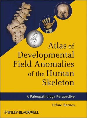
 This item is in your CollectionAtlas of Developmental Field Anomalies of the Human Skeleton: A Paleopathology Perspective
X
 This Item is in Your InventoryAtlas of Developmental Field Anomalies of the Human Skeleton: A Paleopathology Perspective
X
 You must be logged in to review the productsX
 X
 X

Add Atlas of Developmental Field Anomalies of the Human Skeleton: A Paleopathology Perspective, Written by one of the most consulted authorities on the subject, Atlas of Developmental Field Anomalies of the Human Skeleton is the pre-eminent resource for developmental defects of the skeleton. This guide focuses on localized bone structures uti, Atlas of Developmental Field Anomalies of the Human Skeleton: A Paleopathology Perspective to the inventory that you are selling on WonderClubX
 X

Add Atlas of Developmental Field Anomalies of the Human Skeleton: A Paleopathology Perspective, Written by one of the most consulted authorities on the subject, Atlas of Developmental Field Anomalies of the Human Skeleton is the pre-eminent resource for developmental defects of the skeleton. This guide focuses on localized bone structures uti, Atlas of Developmental Field Anomalies of the Human Skeleton: A Paleopathology Perspective to your collection on WonderClub |